October 2006 // Volume 44 // Number 5 // Feature Articles // 5FEA7
An Evaluation of Retinal Imaging Technology for 4-H Beef and Sheep Identification
Abstract
The study reported here evaluated retinal imaging
technology as a means of permanent identification of 4-H beef and sheep.
The OptiReader» Device was used to capture digital images of 491 beef and
220 sheep during 4-H enrollment. A total of 317 beef and 159 sheep were re-imaged.
The on-site visual verification rate was 96.2% for beef and 100% for sheep.
A visual verification exercise showed that individuals could identify a pair
of retinal images as a match 98.6 % of the time for beef. Retinal imaging
is a viable method for enrolling beef and sheep.
Introduction
As the United States government moves towards a National Animal Identification System (United States Animal Identification Plan, 2003), state 4-H programs are seeking new methods of permanent identification for enrolling livestock projects. It is important to verify the identity of animal projects to ensure 4-H members are abiding by possession deadlines and exhibition requirements. Animal verification needs to be a rapid, inexpensive process that is tamper proof.
Solis and Maala, from the University of the Philippines, were the first to characterize the nose printing process in 1975. They realized that identification methods such as branding, tattooing, ear tagging, and ear notching were flawed in that the identifier could be replicated, replaced, or modified, and thus they chose muzzle printing as an alternative. Their findings concluded that nose prints were unique among animals, and the process was a suitable method for identification (Solis & Maala, 1975).
Indiana 4-H beef and sheep projects are currently identified by nose prints and five-digit ear tags (Rusk, 2004, 2004a). Nose prints are used at both the county and state fair levels to verify that the exhibited animal is the same one that was enrolled by an earlier deadline and weigh-in. Nose prints provide a cost effective, real-time verification, but they are inconsistent in quality, sometimes difficult to read due to smearing, and require a "trained eye" to verify a match. Ear tags pose a problem for permanent animal identification because they can be lost or moved from one animal to another (Neary & Yager, 2002). DNA is a reliable form of identification, but it can be costly and can require days or weeks to get results.
An alternative method of animal identification is a biometric, defined as a "measurable physical or behavioral trait used to recognize the identity of an individual" (Elliott, 2003). Iris recognition and retinal imaging are two examples of biometrics that are applicable to animals, and each is present from birth. Dr. Carleton Simon discovered in 1936 that retinal vascular patterns were unique in humans. In 1978, Huntzinger and Christian determined that retinal vascular patterns were also unique between twins. Retinal imaging has been used since the 1970's by the U.S. Navy as a means of secure access.
The ease of obtaining an image of the retinal vascular pattern makes this a logical choice for animal identification. Optibrand, Ltd, LLC, is an animal identification company in Fort Collins, Colorado, specializing in source verification systems. Optibrand has coupled the retinal image with the global positioning satellite system (GPS) to verify the location of the animal when the retinal image is collected (Whittier, Shadduck, & Golden, 2003).
The study reported here evaluated retinal imaging technology and compared it to nose printing as a means of permanent identification for enrolling 4-H beef and sheep projects. Visual verification exercises were administered to Extension educators and adult volunteers to determine false match and false non-match rates for nose printing and retinal imaging. The objectives of the study were to:
-
Compare the time required to obtain a retinal image with the time required to obtain a nose print.
-
Determine the false match and false non-match rates of visual verification of retinal images and nose prints.
The two hypotheses of this study were:
Ho1: Retinal imaging and nose prints are equally viable forms of permanent animal identification.
Ho2: The false match and false non-match rates of retinal images and nose prints are equal.
Methodology
4-H Youth Development educators in the 92 Indiana counties were contacted and given a short explanation about the study. Educators wanting their county to serve as a test site provided the investigator with the times and dates of their beef and sheep enrollment days and the check-in time and date for beef and sheep at the county fair. From the list of volunteer counties, the investigator chose eight Indiana counties to participate in the study.
The youth educators, in the counties selected to participate in the study were sent information about the study and an informed consent form for each 4-H member enrolled in beef and sheep. Participation in this study was voluntary. 4-H members and one of their parents were required to sign an informed consent form prior to images being collected from their animals. In addition to the retinal imaging, county committee members collected nose prints and placed a five-digit tag in the animal's ear.
Image Collection
The OptiReader™ Device, designed by Optibrand Ltd., LLC, was used to capture retinal images from 491 beef and 220 sheep during 4-H enrollment in eight Indiana counties. Prior to attending any county event, a Compact Flash� disk was configured using Optibrand's Reader Configuration software. The researcher selected a one or two eye collection session and then entered categories for the name of the 4-H member(s) enrolling the animal, the animal's ear tag number, and the county of 4-H enrollment.
Retinal imaging required practice for the user to become efficient in collecting images. According to Optibrand, the camera should be held at a 45-degree angle to the eye when imaging cattle and at a slightly flatter angle for sheep. The camera must be held at the proper distance and angle from the eye to prevent blurry images or ones with glare spots. Glare spots that obstruct the retinal pattern in the center of the image are not acceptable; however, glare spots located on the outer edge of the image are tolerable. It is important to keep the camera lens clean and free of dirt and condensation, which can cause interference with obtaining a focused image.
During scanning, the camera was slowly brought towards the animal's eye, keeping the central artery and vein centered on the screen of the OptiReader™ Device. The OptiReader™ Device captured an image once it determined the image was suitable for verification purposes.
The enrollment software recorded the time required to collect a retinal image, which is defined as the moment the targeting is activated until an image is captured that presents the technician with a clear picture of the vein pattern. Nose prints were also collected from participating animals for comparison purposes. The investigator recorded the time from when ink was placed on the nose of an animal until an acceptable nose print had been collected on an index card.
Following collection of retinal images during county enrollment days, the images were converted to jpeg (joint photographic experts group) images from the original blob (binary large object) file and resized to fit a specified frame size within a report. The conversion to the jpeg image allowed the researcher to store the images in a compressed digital format to embed into a Microsoft� Access database file. In addition to the retinal image, the database contained the name(s) of the 4-H member(s) enrolling the animal, the animal's species, visual tag number and breed, along with the enrollment date, the county name, and GPS coordinates. A certificate was created for each animal and printed individually. This report was taken to the county fair and used to compare the original images to the image taken at the fair for on-site, visual verification.
Re-Imaging
A total of 317 beef and 159 sheep were re-imaged at county fairs to compare enrollment and verification images. The verification image was displayed on the screen of the OptiReader™ Device for comparison purposes and then stored on a Compact Flash� disk for inspection at a later time. Several criteria were met before the researchers would declare a set of retinal images a match. The first criterion looked at the basic structure, shape, and number of branches. Due to the angle of the camera, an exact duplicate image cannot be taken, but the main characteristics of the pattern will be present. The relative distance between branches was the second criterion, which allowed for closer inspection of the positions of branches within the pattern. The third criterion included internal structures such as an open space created by the intertwining of branches. See Figure 1 for an example of retinal images.
Figure 1.
Example of Matching Retinal Images
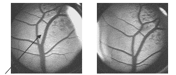
The "kite shaped" opening indicated by the arrow at the center of the images in Figure 1 is an example of internal structures. The distinct shape of the structure along with the definitive branching pattern allows for these images to be declared a match. If a visual match could not be made between two images, the images were submitted to Optibrand Ltd. LLC, who developed computer software to analyze the collected images and verify whether a match existed between the image in question and an image already in the database.
Verification Exercise
A verification exercise was given to 38 4-H Youth educators and adult volunteers involved in the 2004 Animal Sciences Workshop for Youth, to determine the rate of visual verification of retinal images and nose prints for both beef and sheep. The target group simulated the population that would be required to read the retinal images for real-time visual verification in a county fair setting. Volunteers were asked to determine if 20 pairs of retinal images (10 from beef and 10 from sheep) and 20 pairs of nose prints (10 from beef and 10 from sheep) were a match.
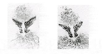
Each participant was given the same image sets in random order and asked to determine if each pair was a match by circling YES, NO, or UNSURE. A short description of retinal images and nose prints was given to the participants to give them background information. The participants were considered to be untrained, however, because specifics were not given as to how to determine if a pair of images was indeed a match. This exercise was divided up into four sections and timed to determine if differences existed in the time it took to visually verify a nose print and a retinal image, and if there was a difference based on species. See Figure 2 for examples of nose print matches in sheep.
Figure 2.
Examples of Matching Nose Prints Taken from Sheep
Data Analysis
Data were entered into Statistical Package for Social Science (SPSS 12.0 for Windows, 2003) and were used to calculate descriptive statistics and make comparisons between species, locations, and methods by two-way Analysis of Variance (ANOVA). The statistics were used to organize and interpret the results of the study and to make recommendations for future uses of the technology.
Results
Table 1 contains a summary of the mean time in seconds required to collect nose prints and retinal images from beef and sheep. The data show no statistical difference in the time required to collect nose prints from beef and sheep projects. It took longer to collect retinal images from both beef and sheep than it took to collect nose prints from these same animals. The data also show that a greater time was required to collect retinal images from sheep than from beef projects.
|
I.D. Method |
N |
Species |
Mean Time (sec) |
|
Nose print |
444 |
Beef |
25.25a |
|
171 |
Sheep |
22.25a |
|
|
Retinal Image |
444 |
Beef |
38.98b |
|
171 |
Sheep |
56.03c |
|
| abc Values in the same row with different superscripts are significantly different (P < .05) | |||
Table 2 shows that method is the most influential factor in this model, and suggests that the mean time needed to collect identification information from each species varies by identification method and the species of the animal being identified. The P-value of <0.001 signifies that at the 95 percent confidence the differences in mean time it took to collect information is due to differences in species and the method of identification, not due to random error. The interaction between method of identification and species is significant.
|
Mean Square |
F-value |
P-value |
|
|
Method |
152519.548 |
100.403 |
<.001 |
|
Species |
13074.886 |
8.607 |
<.001 |
|
Method * Species |
27908.527 |
18.372 |
<.001 |
Table 3 shows the number of animals enrolled in the system at each county, as well as the species and percentages of the images verified, both visually and overall. The overall verified percentage includes those images that were sent to Optibrand for electronic verification (Moss et al., 2004). Of the 474 animals that were re-imaged, a total of 18 retinal images were submitted to technicians at Optibrand for further verification because the researcher was unable to declare them to be matches using visual comparison. An additional eight images were matched by Optibrand technicians using electronic methods.
|
County |
Species |
# Enrolled |
# Re-imaged |
% Verified Visually |
% Verified Overall |
|
Boone |
Bovine |
70 |
45 |
100 |
100 |
|
Elkhart |
Bovine |
163 |
84 |
88.8 |
91.7 |
|
Fountain |
Bovine |
88 |
66 |
100 |
100 |
|
Huntington |
Bovine |
78 |
57 |
91.2 |
96.4 |
|
Porter |
Bovine |
92 |
65 |
95.4 |
98.5 |
|
Adams |
Ovine |
29 |
24 |
100 |
100 |
|
Hancock |
Ovine |
101 |
67 |
100 |
100 |
|
Jasper |
Ovine |
90 |
66 |
100 |
100 |
|
Total |
711 |
474 |
96.3 |
98.7 |
The remaining 10 images were not declared a match because not enough data points overlapped between enrollment and verification images to provide sufficient evidence of being a match. The lowest percentage of visually verified retinal images came from Elkhart County, which was the first county where retinal images were collected by the researcher. It is assumed that the researcher had not yet "mastered" the art of image collection, and thus, some of these early images were not of the quality needed for verification purposes.
The results of the visual verification exercise, shown in Table 4, reveal a 29.7% advantage for correctly identifying retinal images from beef cattle versus nose prints. This same trend was also true for sheep. The percentage of answers recorded as unsure was negligible for retinal images but ranged from 5.26 to 8.42% for nose prints.
|
N |
Overall Percentage Correct |
Percentage of Unsure Answers |
||
|
Beef |
Nose print |
380 |
68.94a |
8.42d |
|
Retinal Image |
370 |
98.64b |
0.27e |
|
|
Sheep |
Nose print |
380 |
79.47c |
5.26d |
|
Retinal Image |
370 |
84.86c |
0.00e |
|
| abcde Values in the same row with different superscripts are significantly different (P < .05) | ||||
The results in Table 5 show a lower rate of false match and false non-match for retinal images than for nose prints from beef. A false match occurred when a participant identified a pair of images as a match, when in fact it was not a match. A false non-match occurred when a participant indicated a pair of images or nose prints were not a match, when in fact they were a match.
|
Correct Response |
True Match |
True Non-Match |
|||||
|
Participant Response |
Match |
False Match |
Unsure |
False Non-Match |
Non-Match |
Unsure |
|
|
Beef |
Nose print |
52.6a |
43.2d |
13.2h |
11.1a |
85.3c |
3.7e |
|
Retina |
99.5b |
0.5e |
0.0i |
1.6b |
97.8d |
0.5f |
|
|
Sheep |
Nose print |
73.7c |
20.5f |
5.8j |
10.0a |
85.3c |
4.7e |
|
Retina |
72.4c |
27.6g |
0.0i |
2.7b |
97.3d |
0.0f |
|
| abcdefghij Values in the same row with different superscripts are significantly different (P < .05) | |||||||
Conclusions
The results of the study reported here lead the researchers to accept hypothesis one: retinal imaging and nose printing are equally reliable forms of permanent identification. The researchers concluded that it takes slightly longer to collect retinal images than to collect nose prints. However, the quality of the image is more important than collection time when verifying the identity of an animal. The study found that untrained individuals were able to correctly identify pairs of retinal images more often than they were able to match nose prints. This implies that county committees and Extension educators can conduct retinal image verifications without hiring a professional to verify the images.
Based on the results of the study, the second hypothesis was rejected. The false match and false non-match rates of visual verification of retinal images were lower than the rates for nose prints. Untrained individuals had higher scores on the exercise to match retinal beef images than they did on sheep images, although there are no structural differences in the retinal vascular patterns of beef and sheep.
In conclusion, the retinal imaging system is a viable method for enrolling beef and sheep projects. Additional research should be conducted to more closely gauge false match and false non-match rates of visual verification and to explore the use of this technology in other species.
Implications
The results of the study reported here have the potential to affect Extension personnel nationwide. Youth livestock specialists and Extension educators involved with county and state fairs across the country are looking for a form of real-time verification to ensure that the animals being exhibited at their county and state shows are the same animals enrolled earlier in the season by their 4-H and FFA members. Likewise, show officials from national livestock shows have similar needs. Many of the national shows are currently requiring exhibitors to submit DNA samples, in the form of either hair or blood, from the animals they plan to exhibit 6 months in the future. Some county and state fairs have similar requirements for the livestock exhibited at their shows, while other fair officials are currently using nose prints to verify animal identity.
As noted earlier, DNA is a reliable form of identification, but it can be costly and usually requires days or weeks to get results. Nose prints provide a cost effective, real-time verification, but they are inconsistent in quality, sometimes difficult to read due to smearing, and require a "trained eye" to verify a match. Retinal images provide the real-time verification feature of nose prints, with the unique permanent identification of DNA. As shown in the study, retinal images are as reliable as nose prints, but do not require a trained individual to verify that two images are the same or different.
In recent years, steers and lambs have been disqualified at state fairs because the original nose print collected at the county level was declared unsatisfactory for matching purposes by the professional print reader hired by the state fair. Retinal images taken with the OptiReader™ device are more consistent in quality than nose prints, because the device itself determines when an image is acceptable. With a laptop computer and the OptiReader™ device, Extension personnel can positively verify sheep and goat projects at ringside, and beef projects at a chute or scale. The real-time verification feature of the retinal imaging process allows show officials to verify animal identity before champions are selected, thus eliminating the awkward and sometimes stressful situations created when champion animals are disqualified after their true identity is determined days and/or weeks following a show.
The only drawbacks of the retinal imaging technology are the cost of the equipment and the time required to learn to operate the OptiReader™ device. In the study reported here, the researcher learned to effectively collect retinal images after only 2 days of training. The cost of the equipment, which includes the necessary software, should be spread over multiple animals and several years of collection in order to make a fair comparison with nose printing. By planning ahead and budgeting for the cost of the retinal scanning equipment, county and state Extension staff can find ways to make this technology affordable. Once purchased, this technology has the potential to be the method of choice for real-time animal verification nationwide.
References
Elliott, S. J. (2003). First working draft--INCITS M1 vocabulary harmonization. Retrieved March 23, 2004 from http://www.incits.org/tc_home/m1htm/docs/m1030356.pdf
Huntzinger, R. S., & Christian, J. C. (1978). The retinal blood vessel patterns in twins. Twin Research: Clinical Studies, 24: 241-246.
Moss, G. E., Whittier, J. C., Stobart, R. H., Baker, D. S., Doubet, J. T., & Golden, B. L. (2004). Computer matching of digital images of retinal vascular patterns of sheep for animal verification. In American Society of Animal Science, Vol. 55. Proceedings of Western Section. Laramie: University of Wyoming.
Neary, M., & Yager, A. (2002). Methods of livestock identification. (Farm Animal Management @ Purdue). Purdue University, Department of Animal Sciences. West Lafayette, Indiana.
Rusk, C. P. (2004). Beef cattle. In Kay Peterson (Ed). 4-H/FFA Handbook/Premium List (pp73-96). Indianapolis, IN: Indiana State Fair.
Rusk, C. P. (2004a). Sheep. In Kay Peterson (Ed). 4-H/FFA Handbook/Premium List (pp137-168). Indianapolis, IN: Indiana State Fair.
Simon, C. (1936, September). The retinal method of identification. Paper presented at the meeting of the International Association of Chief of Police, Kansas City, MO.
Solis, J. A., & Maala, C. P. (1975). Muzzle printing as a method for identification of cattle and carabaos. Philippine Journal of Veterinary Medicine, 14(1): 1-14.
SPSS Version 12.0 for Windows [Computer Software]. (2003). Chicago, IL: SPSS, Inc.
United States Animal Identification Plan. Version 4.1. December 23, 2003. Retrieved January 16, 2004 from http://usaip.info/USAIP4.1.pdf.
Whittier, J., Shadduck, J. A., & Golden, B. L. (2003). Secure identification, source verification of livestock--The value of retinal images and GPS. S. Cox Ed. Precision livestock farming. Wageningen Acad. Pub. The Netherlands. 167-172.